Abstract
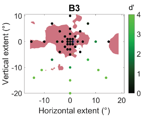
Individuals with macular degeneration typically lose vision in the central region of one or both eyes. A binocular scotoma occurs when vision loss occurs in overlapping locations in both eyes, but stereopsis is impacted even in the non-overlapping region wherever the visual field in either eye is affected. We used a novel stereo-perimetry protocol to measure local stereopsis across the visual field (up to 25° eccentricity) to determine how locations with functional stereopsis relate to the scotomata in the two eyes. Participants included those with monocular or binocular scotomata and age-matched controls with healthy vision. Targets (with or without depth information) were presented on a random dot background. Depth targets had true binocular disparity of 20’ (crossed), whereas non-depth targets were defined by monocular cues such as contrast and dot density. Participants reported target location, and whether it was in depth or flat. Local depth sensitivity (d’) estimates were then combined to generate a stereopsis map. This stereopsis map was compared to the union of the monocular microperimetry estimates that mapped out the functional extent of the scotoma in each eye. The “union” prediction aligned with residual stereopsis, showing impaired stereopsis within this region, and residual stereopsis outside this region. Importantly, the stereo-blind region was typically more extensive than the binocular scotoma defined by the intersection (overlap) of the scotomata. This explains why individuals may have intact binocular visual fields, but be severely compromised in tasks of daily living that benefit from stereopsis, such as eye-hand coordination and navigation.
Journal
Volume
Issue
Year of Publication