- non-photophobic (non-Ph, left panel) patients with severely photophobic (severe-Ph, right panel).")

- Principal Investigator:
- Lora Likova
The purpose of this grant is to identify the mechanisms responsible for generating photophobia in patients who have suffered mild traumatic brain injury (mTBI). Currently, estimates indicate that this painful condition persists in about 60% of those who suffered from blast-related traumatic brain injury and 30% of those who suffered non-blast-related concussive injuries.
We hypothesize that one of two mechanisms -- independently or jointly -- are primarily responsible for the symptoms of photophobia. The first mechanism we propose to test is that a special type of retinal cells, the melanopsin-containing retinal ganglion cells (mRGCs), which project to the brainstem, and/or their pathway, are damaged in TBI. These cells respond to very specific wavelengths of light, so by careful choice of stimuli we can selectively activate their pathway. The second mechanism that we will investigate is the tissue swelling from concussive injury that develops in core brain structures, such as the brainstem.
To investigate these two mechanisms, we propose to test two groups of subjects who have experienced mTBI: one group with photophobia or light-induced pain symptoms, the second group with no photophobia symptoms. We will use a suite of visual diagnostic techniques to determine the differences between these two mTBI groups. We will interpret any systematic differences that we find between these two groups as due to the mechanisms associated with photophobia.
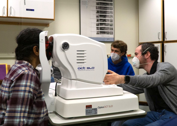
(1) We will measure electrical activity on the surface of the cornea of the eye as a measure of retinal activity of the mRGCs (electroretinography; ERG).
(2) We will record electrical activity from the scalp to determine if different cortical areas are affected by changes in mRGCs (high density electroencephalography; EEG).
(3) We will record changes in the blood oxygenation-level dependent signal in magnetic resonance imaging (MRI) scans of the brain to determine changes in core brain function.
4) We will measure the volume and shape of deep brain structures to determine if there are changes due to tissue swelling using structural MRI techniques, tensor-based morphometry (TBM), in particular.
For those who are experiencing photophobia, it can have a debilitating effect in everyday life. Even normal levels of light can be intensely painful. Depending on the level of photophobia, patients are forced to restrict their activities to many indoor situations or use sunglasses or other aids to reduce the light entering their eyes even when in moderate outdoor lighting. Photophobia is often linked to incessant headaches, insomnia, and irritability. Whether a single of multiple mechanisms cause these disparate symptoms is unknown.
If our efforts to identify the mechanism of photophobia are successful, we will begin the process of finding cures or effective treatments. The major methods to relieve the symptoms temporarily of photophobia are quite limited at present, consisting of dark glasses or colored lenses. Successful completion of our studies will immediately result in improved methods for the objective identification of photophobia. Developing the ability of differential diagnosis will provide the crucially needed scientific, mechanism evaluation that will lead to application of novel treatments within a few years of the completion of our study.
To our best knowledge, no cure has been developed for photophobia to date, as the underlying cause is not yet known. Thus, an effective cure or permanent symptomatic relief will be an invaluable help to those that suffer from photophobia. We expect that there will be substantial long-term beneficial effects from treatments based on the output of the studies we have planned.