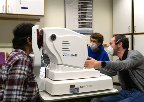
The purpose of this research study is to use the spectral electroretinogram (ERG) to deteremine how the retinal mechanisms of sufferers from abnormal light sensitivity due to head injury differ from those without abnormal light sensitivity.
Home / Projects / Spectral ERG analysis of hypersensitivity to light in traumatic brain injury
Spectral ERG analysis of hypersensitivity to light in traumatic brain injury

Principal Investigator:
Christopher TylerPeople
Get Involved
If you are interested in vision science or want to learn more about low vision and blindness, there are many opportunities to get involved at The Smith-Kettlewell Eye Research Institute.


